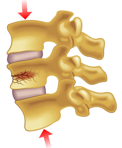
Vertebral Compression Fractures

The type of fracture in the spine that is typically caused by osteoporosis is generally referred to as a compression fracture. A compression fracture is usually defined as a vertebral bone in the spine that has decreased at least 15 to 20% in height due to fracture.
To watch a video about neck strains, click here:
http://www.spine-health.com/video/spinal-compression-fracture-video
These compression fractures can occur in vertebrae anywhere in the spine, but they tend to occur most commonly in the upper back (thoracic spine), particularly in the lower vertebrae of that section of the spine (e.g. T10, T11, T12). They rarely occur above the T7 level of the spine. They often occur in the upper lumbar segments as well, such as L1.
Types of Fracture
A spinal fracture due to osteoporosis (weak bones) is commonly referred to as a compression fracture, but can also be called a vertebral fracture, osteoporotic fracture, or wedge fracture.
The term "wedge fracture" is used because the fracture usually occurs in the front of the vertebra, collapsing the bone in the front of the spine and leaving the back of the same bone unchanged. This process results in a wedge-shaped vertebra. A wedge compression fracture is generally a mechanically stable fracture pattern.
While wedge fractures are the most common type of compression fracture, there are other types as well, such as:
- Crush fracture. If the entire bone breaks, rather than just the front of the vertebra, it may be called a crush fracture.
- Burst fracture. This type of fracture involves some loss of the height in both the front and back walls of the vertebral body (rather than just the front of the vertebra). Making this distinction is important because burst fractures can be unstable and result in progressive deformity or neurologic compromise.
Compression Fracture Symptoms
Vertebral fractures are usually followed by acute back pain, and may lead to chronic pain, deformity (thoracic kyphosis, commonly referred to as a dowager's hump), loss of height, crowding of internal organs, and loss of muscle and aerobic conditioning due to lack of activity and exercise.
A combination of the above problems from vertebral fractures can also lead to changes in the individual's self-image, which in turn can adversely affect self-esteem and ability to carry on the activities of daily living.
Because the majority of damage is limited to the front of the vertebral column, the fracture is usually stable and rarely associated with any nerve or spinal cord damage.
Spinal Fractures are Common
Spinal compression fractures that occur as a result of osteoporosis are actually quite common, occurring in approximately 700,000 people in the U.S. each year.
Osteoporosis is especially common in postmenopausal women. In fact, it is estimated that approximately 25% of all postmenopausal women in the United States have had a vertebral compression fracture.
While osteoporosis is far more prevalent in women - approximately four times as many women have low bone mass or osteoporosis as men - it still occurs in men. As many as 25% of men over age 50 will suffer a bone fracture (e.g. hip or spine) due to osteoporosis.
The problem is that the fracture is not always recognized or accurately diagnosed - instead, the patient's pain is often just thought of as general back pain, such as from a muscle strain or other soft tissue injury, or as a common part of aging. As a result, approximately two thirds of the vertebral fractures that occur each year are not diagnosed and therefore not treated.
TREATMENT: Vertebral Augmentation (or “Kyphoplasty”)
The goals of a kyphoplasty surgical procedure are designed to stop the pain caused by a spinal fracture, to stabilize the bone, and to restore some or all of the lost vertebral body height due to the compression fracture.
To watch a video about vertebroplasty (kyphoplasty without the balloon technology), please click below:
http://www.spine-health.com/video/vertebroplasty-interactive-video
Performing Kyphoplasty Surgery
- During kyphoplasty surgery, a small incision is made in the back through which the doctor places a narrow tube. Using fluoroscopy to guide it to the correct position, the tube creates a path through the back into the fractured area through the pedicle of the involved vertebrae.
- Using X-ray images, the doctor inserts a special balloon through the tube and into the vertebrae, then gently and carefully inflates it. As the balloon inflates, it elevates the fracture, returning the pieces to a more normal position. It also compacts the soft inner bone to create a cavity inside the vertebrae.
- The balloon is removed and the doctor uses specially designed instruments under low pressure to fill the cavity with a cement-like material called polymethylmethacrylate (PMMA). After being injected, the pasty material hardens quickly, stabilizing the bone.
Kyphoplasty surgery to treat a fracture from osteoporosis is performed at a facility under local or general anesthesia. Other logistics for a typical kyphoplasty procedure are:
- The kyphoplasty procedure takes about one hour for each vertebra involved.
- Patients will be observed closely in the recovery room immediately following the kyphoplasty procedure.
- Patients should not drive until they are given approval by their doctor. They will need to arrange for transportation home from the hospital.
Recovery from Kyphoplasty
Pain relief will be immediate for most patients. In others, elimination or reduction of pain is reported within two days. At home, patients can return to their normal daily activities, although strenuous exertion, such as heavy lifting, should be avoided for at least six weeks. Some patients may not experience relief for the first few weeks, but will be followed closely and managed by their physician. Patients should see their physician to begin or review their treatment plan for osteoporosis, including medications to prevent further bone loss.
Candidates for Kyphoplasty
Kyphoplasty cannot correct an established deformity of the spine, and certain patients with osteoporosis are not candidates for this treatment. Patients experiencing painful symptoms or spinal deformities from recent osteoporotic compression fractures are likely candidates for kyphoplasty. The procedure should be completed within 8 weeks of when the fracture occurs for the highest probability of restoring height.
REFERENCES:
http://www.spine-health.com/conditions/osteoporosis/when-back-pain-a-spine-compression-fracture


